Scientists Clarify How a Brain Protein Linked to Mental Health Conditions Functions in Developing Memory Circuits
Results refine scientific models of how brain cells build communication networks

Scientists at the UC Irvine Center for the Neurobiology of Learning and Memory (CNLM) have published new research that clarifies how a brain protein connected to several mental health disorders behaves in developing memory circuits. The findings help resolve uncertainty about how strongly this protein shapes communication between brain cells.
The study focuses on MDGA1, a protein found on the outer surface of neurons. Human genetic studies have linked MDGA1 to conditions such as schizophrenia, bipolar disorder, and depression. Because these disorders often involve changes in how neurons connect and signal to one another, researchers have suspected that MDGA1 might play a key role at synapses, the junctions where brain cells exchange information.
To investigate this, the research team built a new mouse model that allowed them to see exactly where the natural MDGA1 protein appears in the brain and when it is most active during development. They focused on the hippocampus, a region essential for learning and memory.

They found that MDGA1 is most abundant early in brain development and is mainly located along dendrites, the branching parts of neurons that receive incoming signals. Surprisingly, the protein was not highly concentrated at synapses themselves. The team then tested what happens when MDGA1 is selectively removed from specific neurons. Using precise genetic tools and electrical recordings, they measured how well synapses continued to function. In the circuits they studied, loss of MDGA1 did not significantly change excitatory or inhibitory signaling.
“Neurons communicate with one another at synapses. There are trillions of synapses in the human brain, and the correct establishment of each of these connections during development is essential for healthy brain function,” says Dr. Javier Diaz Alonso. “A family of proteins called cell adhesion molecules regulates the formation of synapses, ensuring that only connections between the right partners are formed. For the past decade, researchers have been interested in an unusual member of this family, called MDGA1. These studies were done in vitro (in a dish), because the right tools to visualize MDGA1 in the brain were not available. Our study overcame this limitation, allowing precise identification of MDGA1 in the mouse brain. Surprisingly, we found that, in a region of the hippocampus involved in memory formation, MDGA1 was not enriched at synapses. Even more surprisingly, when we removed MDGA1 from hippocampal neurons, synaptic transmission was largely normal.”

These results suggest that MDGA1 is present during key developmental periods but is not a major driver of synaptic signaling in these memory circuits. That gives researchers a more precise understanding of where this protein fits (and where it does not) in the synapse-building process. This matters because many genes linked to mental health conditions affect how neurons connect and communicate. Designing better treatments depends on having accurate models of how these molecules actually function in brain circuits. By showing where MDGA1 is active, and where it is not, this research helps focus future studies on the mechanisms most likely to influence brain network development and mental health outcomes.
"In the fast-paced world of neuroscience, so-called ‘negative’ results sometimes feel like failure. In many cases, however, such findings pave clearer roadmaps by which to elucidate mechanisms underlying complex diseases. With this study, we are now much closer to understanding when and where MDGA1 may affect neuronal, hippocampal, and brain function, broadly, in neuropsychiatric disease states,” said Matthew Sandoval, a CNLM doctoral student and NIH-funded Schneiderman Training Program in Learning and Memory T32 Scholar. The research was conducted in the laboratory of Dr. Javier Diaz-Alonso, a CNLM Fellow and assistant professor in the UC Irvine Department of Anatomy and Neurobiology who studies how synapses are formed and regulated at the molecular level.
The research was published February 11 in The Journal of Neuroscience. An image from the study was selected for the journal’s front cover (below).
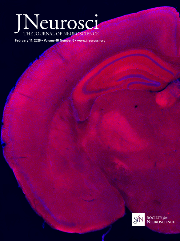
From JNeurosci
"Microscopic image of a coronal brain slice from a juvenile mouse. Shown is expression of a protein associated with schizophrenia and depression (MAM domain-containing glycosylphosphatidylinositol anchor protein 1 or MDGA1, red) with all cells highlighted via a DAPI stain (blue). MDGA1 is visible in the cortex, hippocampus, thalamus, and habenula."
About the Center for the Neurobiology of Learning and Memory
Established in 1983 by the UC Regents, with James L. McGaugh as its Founding Director, the CNLM is the first research institute in the world dedicated to the interdisciplinary study of the fundamental brain mechanisms of learning and memory. It is credited with numerous seminal discoveries about how memory works and is recognized as a global leader in the area. Led by Director Michael Yassa, the CNLM is home to more than 120 faculty scientists at UCI and beyond. The Center’s Office of Outreach and Education develops and leads innovative neuroscience education programs that inspire and train the next generation of neuroscience leaders. For more information, visit cnlm.uci.edu.
For media inquiries, please contact (949) 824-5193 or manuella.yassa@uci.edu